Glioblastoma Radiology Discussion
Author: Jared Nelson, MD
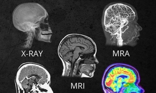
Radiology imaging is essential in the diagnosis, treatment, and surveillance for patients with masses, especially glioblastomas. While useful in so many other areas, x-ray and ultrasound have no significant role to play in glioblastoma imaging.
Computed Tomography (CT) imaging is often the first imaging tool used to first diagnose and intracranial mass, look for mass effect, or accompanying hemorrhage. CT of the head is quick, and does a good job of visualizing those changes, but is insensitive to see the glioblastoma tumor itself.
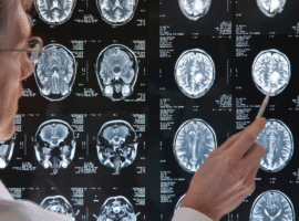
For GBM, magnetic resonance imaging (MRI) is the gold standard to characterize a brain mass, as well as it’s location, and extent of edema and mass effect. This is especially helpful when one is evaluating for response to treatment, e.g., post-operative imaging.
PET imaging depends upon the different degrees to which body tissues and tumors use the radioactive sugar-like substances. Because the brain uses so much of this radiotracer, the background activity of the brain is high and it is hard to separate out the normal brain tissue from the glioblastoma.
-Jared Nelson, MD
Related Post
Glioblastoma vs Astrocytoma: A Patient Guide